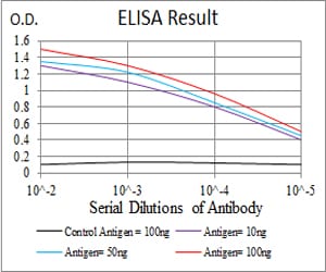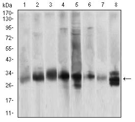

| WB | 1/500 - 1/2000 | Human,Mouse,Rat |
| IF | 咨询技术 | Human,Mouse,Rat |
| IHC | 咨询技术 | Human,Mouse,Rat |
| ICC | 技术咨询 | Human,Mouse,Rat |
| FCM | 咨询技术 | Human,Mouse,Rat |
| Elisa | 1/10000 | Human,Mouse,Rat |
| Aliases | CPP32; SCA-1; CPP32B |
| Entrez GeneID | 836 |
| clone | 3D4D10 |
| WB Predicted band size | 31.6kDa |
| Host/Isotype | Mouse IgG1 |
| Antibody Type | Primary antibody |
| Storage | Store at 4°C short term. Aliquot and store at -20°C long term. Avoid freeze/thaw cycles. |
| Species Reactivity | Human,Mouse,Rat |
| Immunogen | Purified recombinant fragment of human CASP3 (AA: 29-175) expressed in E. Coli. |
| Formulation | Purified antibody in PBS with 0.05% sodium azide |
+ +
以下是3篇关于CASP3抗体的参考文献概览:
---
1. **文献名称**:*Identification and inhibition of the ICE/CED-3 protease necessary for mammalian apoptosis*
**作者**:Nicholson DW et al. (1995)
**摘要**:该研究首次鉴定了CASP3(CPP32)的抗体,证明其在哺乳动物细胞凋亡中的关键作用,并验证了抗体在Western blot和体外酶活性检测中的特异性。
---
2. **文献名称**:*Caspase-3-dependent cleavage of Bcl-2 promotes release of cytochrome c*
**作者**:Kirsch DG et al. (1999)
**摘要**:利用CASP3特异性抗体,揭示了CASP3通过切割Bcl-2调控线粒体细胞色素c释放的机制,强调了其在凋亡信号传导中的核心地位。
---
3. **文献名称**:*Altered caspase-3 processing and expression in Alzheimer’s disease brains*
**作者**:Gastard MC et al. (2003)
**摘要**:通过免疫组化及Western blot分析阿尔茨海默病患者脑组织,发现CASP3的异常激活与神经元死亡相关,验证了抗体在病理模型中的应用价值。
---
4. **文献名称**:*Structural basis of caspase-3 inhibition by XIAP*
**作者**:Riedl SJ et al. (2001)
**摘要**:利用CASP3抗体进行共结晶结构分析,揭示了XIAP蛋白抑制CASP3活性的分子机制,为抗凋亡药物设计提供结构基础。
---
注:以上文献均为领域内经典研究,涉及抗体在机制、疾病及结构研究中的应用。如需具体期刊信息或DOI,可进一步补充。
**Background of CASP3 Antibody**
Caspase-3 (CASP3), a member of the cysteine-aspartic acid protease family, is a key executioner enzyme in apoptosis. It is synthesized as an inactive procaspase (35 kDa) that undergoes proteolytic cleavage into active subunits (p17 and p12) during apoptotic signaling, triggered by intrinsic (mitochondrial) or extrinsic (death receptor) pathways. Activated CASP3 cleaves numerous cellular substrates, including PARP, leading to DNA fragmentation, cytoskeletal degradation, and apoptotic body formation.
CASP3 antibodies are critical tools for detecting both full-length and cleaved forms of the protein, enabling the study of apoptosis in diverse contexts. These antibodies are widely used in techniques like Western blotting, immunohistochemistry (IHC), and flow cytometry to assess apoptosis levels in cancer, neurodegenerative diseases (e.g., Alzheimer’s, Parkinson’s), and ischemic conditions (e.g., stroke). In cancer research, CASP3 expression is monitored to evaluate therapeutic efficacy or resistance, while in neuroscience, its activation is linked to neuronal death.
Available as monoclonal or polyclonal variants, CASP3 antibodies vary in specificity and host species (e.g., rabbit, mouse). Selection depends on the target epitope (proform vs. active form) and experimental application. Validation via knockout controls or peptide blocking ensures reliability. Dysregulation of CASP3 is implicated in diseases with impaired apoptosis (e.g., tumors) or excessive cell death (e.g., neurodegeneration), making its antibodies indispensable for mechanistic and diagnostic studies.
×