
| WB | 咨询技术 | Human,Mouse,Rat |
| IF | 咨询技术 | Human,Mouse,Rat |
| IHC | 咨询技术 | Human,Mouse,Rat |
| ICC | 技术咨询 | Human,Mouse,Rat |
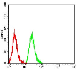
| FCM | 1/200 - 1/400 | Human,Mouse,Rat |
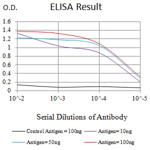
| Elisa | 1/10000 | Human,Mouse,Rat |
| Aliases | LC3; LC3A; ATG8E; MAP1ALC3; MAP1BLC3 |
| Entrez GeneID | 84557 |
| clone | 7E9E12 |
| WB Predicted band size | 14.3kDa |
| Host/Isotype | Mouse IgG2a |
| Antibody Type | Primary antibody |
| Storage | Store at 4°C short term. Aliquot and store at -20°C long term. Avoid freeze/thaw cycles. |
| Species Reactivity | Human |
| Immunogen | Purified recombinant fragment of human MAP1LC3A (AA: 1-121) expressed in E. Coli. |
| Formulation | Purified antibody in PBS with 0.05% sodium azide |
+ +
以下是关于MAP1LC3A抗体的3篇参考文献,按作者和内容概括整理:
---
1. **文献名称**:*"LC3. a mammalian homologue of yeast Apg8p, is localized in autophagosome membranes after processing"*
**作者**:Kabeya et al. (2000)
**摘要**:该研究首次描述了哺乳动物LC3蛋白(包括MAP1LC3A)在自噬过程中的作用。作者开发了针对LC3的特异性抗体,证实LC3-I(胞质形式)向LC3-II(自噬体膜结合形式)的转化是自噬活性的标志,为后续自噬检测提供了关键工具。
2. **文献名称**:*"Methods to Monitor Autophagy Using LC3 Antibodies"*
**作者**:Mizushima et al. (2010)
**摘要**:这篇方法学论文系统评估了LC3抗体(包括MAP1LC3A)在Western blot和免疫荧光中的应用。作者强调了抗体特异性验证的重要性,并比较了不同实验条件下LC3-I/II的检测标准,为自噬研究提供了实用指南。
3. **文献名称**:*"Differential expression patterns of LC3A and LC3B in human cancer cell lines"*
**作者**:Koukourakis et al. (2012)
**摘要**:研究通过MAP1LC3A和LC3B特异性抗体,分析了多种癌细胞系中LC3同工型的表达差异。结果显示LC3A在肿瘤细胞的自噬和应激反应中具有独特分布,提示其可能作为特定癌症类型的生物标志物。
---
如需更多文献或具体应用场景(如神经疾病、免疫印迹优化),可进一步补充关键词检索。
MAP1LC3A (microtubule-associated protein 1 light chain 3 alpha), also known as LC3A, is a key autophagy-related protein involved in the formation and maturation of autophagosomes—double-membrane vesicles that degrade cellular components during autophagy. As a mammalian homolog of yeast Atg8. LC3A exists in two forms: cytosolic LC3-I and lipidated, membrane-bound LC3-II, the latter serving as a hallmark of autophagic activity. Antibodies targeting MAP1LC3A are widely used to monitor autophagy flux in research, as the conversion of LC3-I to LC3-II correlates with autophagosome formation.
These antibodies are essential tools in studying autophagy's role in physiological processes (e.g., nutrient deprivation, cellular stress) and diseases such as cancer, neurodegeneration, and infectious diseases. They enable detection via techniques like Western blotting, immunofluorescence, and immunohistochemistry, helping visualize punctate LC3-II structures in cells. Specificity for LC3A is critical due to the existence of paralogs (LC3B, LC3C) with overlapping functions. Researchers often validate results using autophagy inhibitors (e.g., bafilomycin A1) or inducers (e.g., rapamycin) to confirm dynamic changes.
Dysregulation of LC3A-mediated autophagy is linked to pathological conditions, making its antibody a valuable asset in both mechanistic studies and therapeutic exploration targeting autophagy pathways.
×