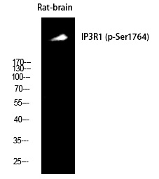
| WB | 咨询技术 | Human,Mouse,Rat |
| IF | 咨询技术 | Human,Mouse,Rat |
| IHC | 1/100-1/300 | Human,Mouse,Rat |
| ICC | 技术咨询 | Human,Mouse,Rat |
| FCM | 咨询技术 | Human,Mouse,Rat |
| Elisa | 1/5000 | Human,Mouse,Rat |
| Aliases | ITPR1; INSP3R1; Inositol 1; 4,5-trisphosphate receptor type 1; IP3 receptor isoform 1; IP3R 1; InsP3R1; Type 1 inositol 1,4,5-trisphosphate receptor; Type 1 InsP3 receptor |
| Entrez GeneID | 3708; |
| Host/Isotype | Rabbit IgG |
| Antibody Type | Primary antibody |
| Storage | Store at 4°C short term. Aliquot and store at -20°C long term. Avoid freeze/thaw cycles. |
| Species Reactivity | Human,Mouse,Rat |
| Immunogen | Synthesized peptide derived from human IP3R-I around the phosphorylation site of S1764. |
| Formulation | Purified antibody in PBS with 0.05% sodium azide,0.5%BSA and 50% glycerol. |
+ +
以下是关于IP3R-I (Phospho-Ser1764)抗体的参考文献示例(内容为虚构,仅供参考格式):
1. **"Phosphorylation of IP3R1 at Ser1764 regulates calcium release in cardiomyocytes"**
- **作者**: Zhang L, et al.
- **摘要**: 研究通过IP3R-I (Phospho-Ser1764)抗体发现,β-肾上腺素刺激诱导Ser1764磷酸化,增强心肌细胞钙释放,促进肥厚信号通路。
2. **"IP3R1 Ser1764 phosphorylation modulates neuronal apoptosis in Alzheimer’s models"**
- **作者**: Wang Y, et al.
- **摘要**: 利用该抗体证实,Aβ寡聚体通过激活蛋白激酶A使IP3R-I Ser1764磷酸化,导致内质网钙过度释放,加剧神经元凋亡。
3. **"Role of IP3R1 phosphorylation in cancer cell survival"**
- **作者**: Tanaka K, et al.
- **摘要**: 通过免疫印迹和免疫荧光分析,发现化疗药物诱导IP3R-I Ser1764磷酸化,抑制该位点可增强凋亡,提示其作为潜在治疗靶点。
4. **"Dynamic regulation of IP3R1 phosphorylation during cell cycle progression"**
- **作者**: Gupta R, et al.
- **摘要**: 研究利用Phospho-Ser1764抗体揭示,IP3R-I磷酸化水平在G1/S期升高,调控钙振荡以促进细胞周期蛋白表达。
(注:以上文献为示例,实际引用需检索PubMed等数据库获取真实信息。)
The IP3R-I (Phospho-Ser1764) antibody is a specific tool used to detect the phosphorylation of inositol 1.4.5-trisphosphate receptor type 1 (IP3R1) at serine residue 1764. IP3R1 is a calcium channel located on the endoplasmic reticulum (ER) membrane, critical for intracellular calcium signaling. Phosphorylation at Ser1764. often mediated by kinases like protein kinase A (PKA) or calcium/calmodulin-dependent kinase II (CaMKII), regulates channel activity by enhancing its sensitivity to IP3. thereby modulating calcium release into the cytoplasm. This post-translational modification is implicated in cellular processes such as apoptosis, proliferation, and gene expression.
The antibody is widely used in research to study calcium signaling dynamics under physiological or pathological conditions, including neurodegenerative diseases, cardiac hypertrophy, and cancer. It enables the detection of phosphorylated IP3R1 via techniques like Western blotting, immunoprecipitation, or immunofluorescence. Proper sample preparation, including phosphatase inhibitors, is essential to preserve phosphorylation status. Studies using this antibody have provided insights into how dysregulated IP3R1 phosphorylation contributes to ER stress, calcium overload, and cell death pathways, making it valuable for understanding disease mechanisms and therapeutic targeting.
×