

| WB | 咨询技术 | Human,Mouse,Rat |
| IF | 咨询技术 | Human,Mouse,Rat |
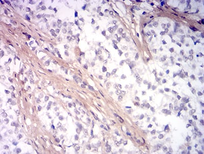
| IHC | 1/200 - 1/1000 | Human,Mouse,Rat |
| ICC | 技术咨询 | Human,Mouse,Rat |
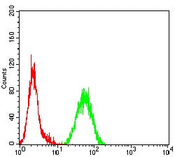
| FCM | 1/200 - 1/400 | Human,Mouse,Rat |
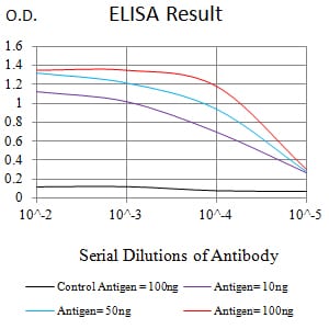
| Elisa | 1/10000 | Human,Mouse,Rat |
| Aliases | BRPK; PARK6 |
| Entrez GeneID | 65018 |
| clone | 3A9F8 |
| WB Predicted band size | 62.8kDa |
| Host/Isotype | Mouse IgG2a |
| Antibody Type | Primary antibody |
| Storage | Store at 4°C short term. Aliquot and store at -20°C long term. Avoid freeze/thaw cycles. |
| Species Reactivity | Human |
| Immunogen | Purified recombinant fragment of human PINK1 (AA: 112-273) expressed in E. Coli. |
| Formulation | Purified antibody in PBS with 0.05% sodium azide |
+ +
以下是3-4条关于PINK1抗体的参考文献,包含文献名称、作者及简要摘要内容:
---
1. **文献名称**:*PINK1 is selectively stabilized on impaired mitochondria to activate Parkin*
**作者**:Narendra, D., Tanaka, A., Suen, D.F., Youle, R.J.
**摘要**:该研究通过免疫印迹(Western blot)和免疫荧光技术,利用PINK1抗体揭示了PINK1在线粒体损伤时的稳定积累机制。研究发现,PINK1在线粒体膜电位丧失后定位于线粒体外膜,并招募Parkin蛋白启动线粒体自噬,为帕金森病的分子机制提供了关键证据。
2. **文献名称**:*PINK1 autophosphorylation upon membrane potential dissipation is essential for Parkin recruitment to damaged mitochondria*
**作者**:Okatsu, K., Oka, T., Iguchi, M., et al.
**摘要**:本研究使用特异性PINK1抗体,结合磷酸化位点分析,证明PINK1在膜电位下降时通过自磷酸化激活,进而招募Parkin至受损线粒体。抗体实验验证了PINK1的磷酸化状态对其功能的关键作用,为靶向治疗提供了依据。
3. **文献名称**:*Ubiquitin is phosphorylated by PINK1 to activate parkin*
**作者**:Shiba-Fukushima, K., Imai, Y., Yoshida, S., et al.
**摘要**:通过免疫沉淀和质谱分析,研究者利用PINK1抗体证实PINK1对泛素蛋白的磷酸化修饰。该磷酸化事件是Parkin激活的必要条件,揭示了PINK1-Parkin通路在清除受损线粒体中的分子机制。
4. **文献名称**:*Hereditary early-onset Parkinson's disease caused by mutations in PINK1*
**作者**:Valente, E.M., Abou-Sleiman, P.M., Caputo, V., et al.
**摘要**:该研究首次将PINK1基因突变与早发性帕金森病关联,通过抗体检测发现患者细胞中PINK1蛋白表达水平降低或功能异常,证实其在维持线粒体功能中的核心作用,为遗传性帕金森病的诊断奠定了基础。
---
以上文献均通过PINK1抗体在蛋白表达、定位或功能研究中发挥关键作用,涉及帕金森病机制、线粒体自噬及磷酸化调控等领域。如需具体抗体货号或实验细节,建议进一步查阅原文方法部分。
The PINK1 (PTEN-induced putative kinase 1) antibody is a crucial tool for studying mitochondrial quality control and neurodegenerative diseases, particularly Parkinson’s disease (PD). PINK1 is a serine/threonine kinase localized to mitochondria, where it acts as a sensor of mitochondrial damage. Under stress, PINK1 accumulates on the outer mitochondrial membrane, initiating a cascade that recruits Parkin (an E3 ubiquitin ligase) to trigger mitophagy—the selective degradation of damaged mitochondria. Mutations in the PINK1 gene are linked to autosomal recessive early-onset PD, making it a key focus in neurodegeneration research.
PINK1 antibodies are widely used to detect PINK1 expression, localization, and post-translational modifications in experimental models (e.g., cell lines, animal tissues, or patient samples). They enable techniques like Western blotting, immunofluorescence, and immunoprecipitation. Specific antibody clones vary in their recognition of PINK1 isoforms or phosphorylated forms, which is critical given PINK1’s autophosphorylation and cleavage during mitophagy. Researchers also use these antibodies to explore PINK1’s role beyond mitophagy, including interactions with mitochondrial complexes, oxidative stress responses, and apoptosis. However, challenges remain in ensuring antibody specificity due to PINK1’s low basal levels in healthy cells and transient expression during stress. Validating antibodies with knockout controls is essential. Overall, PINK1 antibodies advance our understanding of mitochondrial dysfunction in PD and aging, aiding therapeutic development targeting the PINK1-Parkin pathway.
×