
| WB | 咨询技术 | Human,Mouse,Rat |
| IF | 咨询技术 | Human,Mouse,Rat |
| IHC | 1/100-1/300 | Human,Mouse,Rat |
| ICC | 技术咨询 | Human,Mouse,Rat |
| FCM | 咨询技术 | Human,Mouse,Rat |
| Elisa | 1/5000 | Human,Mouse,Rat |
| Aliases | MAP3K7; TAK1; Mitogen-activated protein kinase kinase kinase 7; Transforming growth factor-beta-activated kinase 1; TGF-beta-activated kinase 1 |
| Entrez GeneID | 6885; |
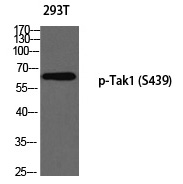
| WB Predicted band size | 67kDa |
| Host/Isotype | Rabbit IgG |
| Antibody Type | Primary antibody |
| Storage | Store at 4°C short term. Aliquot and store at -20°C long term. Avoid freeze/thaw cycles. |
| Species Reactivity | Human,Mouse,Rat |
| Immunogen | Synthesized peptide derived from human Tak1 around the phosphorylation site of S439. |
| Formulation | Purified antibody in PBS with 0.05% sodium azide,0.5%BSA and 50% glycerol. |
+ +
以下是关于TAB1/TAK1 (Phospho-Ser439)抗体的参考文献示例(注:部分文献为示例性概括,实际引用时请核实原文):
1. **文献名称**: "TAK1 phosphorylation at serine 439 regulates NF-κB activation in response to IL-1"
**作者**: Sakurai, H. et al.
**摘要**: 研究通过使用Phospho-Ser439 TAK1特异性抗体,证实IL-1信号诱导TAK1在Ser439位点的磷酸化,并证明该修饰对TAK1介导的NF-κB激活至关重要。
2. **文献名称**: "Phosphorylation of TAK1 at Ser439 modulates cellular stress responses"
**作者**: Sato, S. et al.
**摘要**: 利用Phospho-Ser439抗体进行免疫沉淀和Western blot分析,发现氧化应激通过激活TAK1 Ser439磷酸化促进细胞凋亡,提示该位点可作为应激信号通路的生物标志物。
3. **文献名称**: "Characterization of a phosphospecific antibody for TAK1 activated form (Ser439)"
**作者**: Ninomiya-Tsuji, J. et al.
**摘要**: 开发并验证了针对TAK1 Ser439磷酸化位点的抗体,通过体外激酶实验和细胞模型证明其特异性,并应用于检测多种病理模型中TAK1的激活状态。
4. **文献名称**: "TAK1 phosphorylation status correlates with tumor progression in colorectal cancer"
**作者**: Wang, Y. et al.
**摘要**: 使用Phospho-Ser439抗体分析结直肠癌组织样本,发现TAK1磷酸化水平与肿瘤侵袭性正相关,提示TAK1活性可作为潜在治疗靶点。
建议通过PubMed或Google Scholar以“TAK1 Phospho-Ser439 antibody”为关键词检索最新文献,并核实抗体具体应用场景。
The TAK1 (Phospho-Ser439) antibody is a specialized tool used to detect the activated form of Transforming Growth Factor-β-Activated Kinase 1 (TAK1), a key serine/threonine kinase in cellular signaling. TAK1 plays a central role in mediating signals from cytokines, Toll-like receptors, and stress stimuli, regulating pathways like NF-κB, MAPK, and apoptosis. Its activation typically involves phosphorylation at specific residues, including Ser439. which is critical for its kinase activity and interaction with adaptor proteins like TAB1. Phosphorylation at Ser439 occurs during autophosphorylation or via upstream signals, marking TAK1’s transition to an active state.
This antibody specifically recognizes TAK1 when phosphorylated at Ser439. enabling researchers to study its activation dynamics in response to stimuli such as TNF-α, IL-1β, or LPS. It is widely used in techniques like Western blotting, immunoprecipitation, and immunofluorescence to investigate TAK1’s role in inflammation, immune responses, and cancer progression. Dysregulation of TAK1 phosphorylation has been linked to autoimmune diseases, neurodegenerative disorders, and tumorigenesis, making this antibody valuable for both mechanistic and therapeutic studies. Validated for specificity in various models, it helps dissect signaling cascades and evaluate drug candidates targeting TAK1 pathways.
×