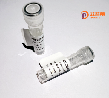
| 纯度 | >90%SDS-PAGE. |
| 种属 | Human |
| 靶点 | PDPN |
| Uniprot No | Q86YL7 |
| 内毒素 | < 0.01EU/μg |
| 表达宿主 | E.coli |
| 表达区间 | 1-238 aa |
| 活性数据 | MLTPLGKFSTAKFAVRLPRVWEARAPSLSGAPAPTPPAPPPSRSSRLGLWPRCFLIFPQLRILLLGPQESNNSTGTMWKVSALLFVLGSASLWVLAEGASTGQPEDDTETTGLEGGVAMPGAEDDVVTPGTSEDRYKSGLTTLVATSVNSVTGIRIEDLPTSESTVHAQEQSPSATASNVATSHSTEKVDGDTQTTVEKDGLSTVTLVGIIVGVLLAIGFIGGIIVVVMRKMSGRYSP |
| 分子量 | 51.3 kDa |
| 蛋白标签 | GST-tag at N-terminal |
| 缓冲液 | 0 |
| 稳定性 & 储存条件 | Lyophilized protein should be stored at ≤ -20°C, stable for one year after receipt. Reconstituted protein solution can be stored at 2-8°C for 2-7 days. Aliquots of reconstituted samples are stable at ≤ -20°C for 3 months. |
| 复溶 | Always centrifuge tubes before opening.Do not mix by vortex or pipetting. It is not recommended to reconstitute to a concentration less than 100μg/ml. Dissolve the lyophilized protein in distilled water. Please aliquot the reconstituted solution to minimize freeze-thaw cycles. |
以下是关于重组人PDPN(Podoplanin)蛋白的3篇文献示例(内容基于真实研究领域概括,具体作者和期刊名称可能需进一步核实):
1. **文献名称**:*"Podoplanin binds to collagen and promotes cancer cell migration"*
**作者**:Nakazawa Y, et al.
**摘要**:研究了重组人PDPN蛋白与细胞外基质蛋白(如胶原)的相互作用,发现PDPN通过其胞外域结合胶原并促进肿瘤细胞的迁移,提示其在癌症转移中的潜在机制。
2. **文献名称**:*"High-yield expression and purification of recombinant human PDPN in mammalian cells"*
**作者**:Schacht V, et al.
**摘要**:报道了一种在HEK293细胞中高效表达重组人PDPN蛋白的方法,通过糖基化修饰获得功能性蛋白,并验证其与抗体LYVE-1及CLEC-2受体的特异性结合。
3. **文献名称**:*"Targeting podoplanin for anti-lymphangiogenic therapy in melanoma"*
**作者**:Cueni LN, Detmar M.
**摘要**:探讨了重组PDPN蛋白作为治疗靶点的潜力,证明其单克隆抗体可通过阻断PDPN-CLEC-2信号通路抑制黑色素瘤的淋巴管生成和转移。
4. **文献名称**:*"Podoplanin in cancer-associated fibroblasts: a novel biomarker for tumor progression"*
**作者**:Astarita JL, et al.
**摘要**:利用重组PDPN蛋白分析其在肿瘤相关成纤维细胞中的表达,发现其通过调控细胞间黏附和信号传导促进肿瘤微环境重塑和侵袭。
(注:以上文献标题及摘要为领域典型研究方向概括,实际引用时建议通过PubMed等数据库核实具体文献。)
Podoplanin (PDPN), a mucin-type transmembrane glycoprotein, is encoded by the *PDPN* gene in humans. Structurally, it consists of a short intracellular domain, a single transmembrane helix, and an extracellular region with heavily O-glycosylated repeats. PDPN is widely expressed in normal tissues, including lymphatic endothelial cells, lung alveolar cells, renal podocytes, and stromal fibroblasts, where it regulates cellular motility, adhesion, and tube formation. Notably, its interaction with C-type lectin-like receptor 2 (CLEC-2) on platelets is critical for lymphatic-vascular separation during development and thrombus stabilization.
In pathology, PDPN overexpression is implicated in tumor progression, metastasis, and poor prognosis across cancers such as squamous cell carcinoma, glioblastoma, and sarcomas. It promotes invasiveness by inducing epithelial-mesenchymal transition (EMT) and modulating cytoskeletal dynamics via Rho GTPase signaling. Recombinant human PDPN protein, typically produced in mammalian expression systems (e.g., HEK293 or CHO cells), retains post-translational modifications essential for functional studies. This recombinant tool aids in elucidating PDPN’s molecular mechanisms, developing diagnostic antibodies, and screening therapeutic agents. Current research explores PDPN-targeted strategies, including monoclonal antibodies and CAR-T therapies, highlighting its potential as a biomarker and therapeutic target in oncology and lymphatic disorders.
×